

July 2019
Accessory Pathway Location
Dr Heather Edwards
ST5 Cardiology SpR at University Hospital Wales
h.edwards24@googlemail.com
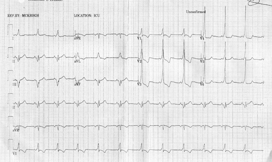
Figure 1
12 lead ECG from a patient with pre-excitation
QUESTION
Where is the location of the accessory pathway?
Answer
Left posterior/Left posterolateral
Explanation
The following can help localise the site of an accessory pathway from a pre-excited ECG:
1. If the initial 20ms of the delta wave in lead I is negative or isoelectric or if the R wave is > than S wave in V1 there is a left sided pathway. If aVF is positive the pathway is lateral/anterolateral. If negative or isoelectric the pathway is posterior or posterolateral.
2. If lead II is negative a subepicardial pathway is likely.
3. If V1 is negative or isoelectric the pathway is septal. If avf is positive and lead III has a dominant R wave the pathway is anteroseptal/right anterior paraseptal. If the S wave is dominant then suspect mid-septal tricuspid annulus. If aVF is –ve then posteroseptal tricuspid annulus or coronary sinus ostium. If aVF is isoelectric the pathway is either posteroseptal mitral or tricuspid annulus.
4. If V1 is positive but not fulfilling step 1 criteria the pathway is right free wall. If aVF is positive the pathway is either right anterior or anterolateral. If aVF is negative or isoelctric and lead II is positive the pathway is right lateral. If lead II is isoelectric the pathways is right posterior or posterolateral.
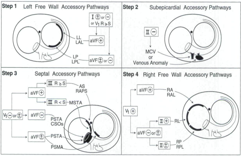
Figure 2
Development and validation of an ECG algorithm for identifying accessory pathway ablation site in Wolff-Parkinson-White syndrome. Arruda et al. J Cardiovasc Electrophysiol. 1998 Jan;9(1):2-12.