March 2024

Variable QRS morphology during a physiological pacing implant

Lloyd Tudor, Specialist Cardiac Physiologist, Leeds Teaching Hospitals NHS Trust
Disclosure: The author has no conflict of interests to declare.

Background

During a physiological pacing (Left Bundle Branch Area Pacing) implant, a dual chamber pacemaker was inserted with an atrial lead in the right atrial appendage and a ventricular lead in the right ventricular septum with the intention of capturing the cardiac conduction system.

A ventricular threshold test was performed and it was noted that the morphology of the paced QRS complex changed during the test prior to a loss of ventricular capture. The test was conducted in VVI mode, decrementing from an output of 5.0 V. The change in QRS morphology occurred at an output of 2.5 V remaining this way until capture was lost at 0.5 V. The QRS duration also changed from 122 ms to 109 ms at this time.

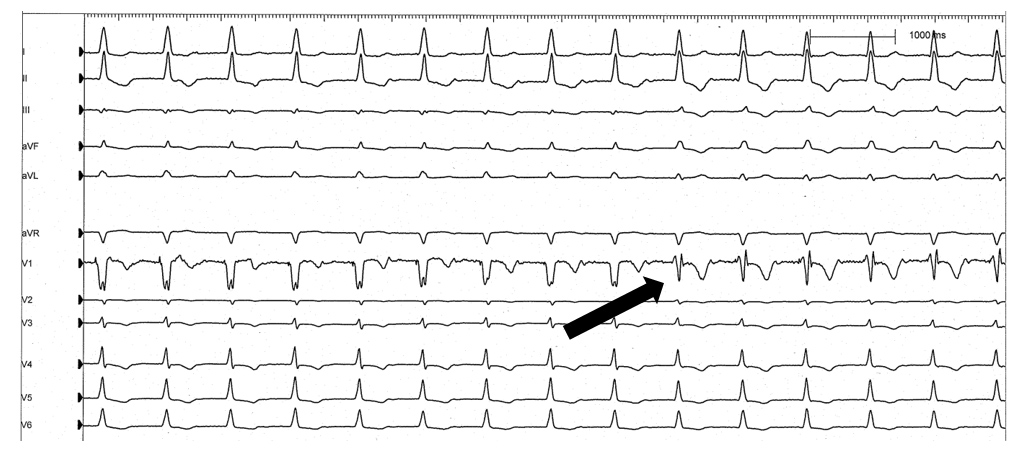
Figure 1. ECG of the ventricular threshold test with the paced QRS morphology change highlighted.

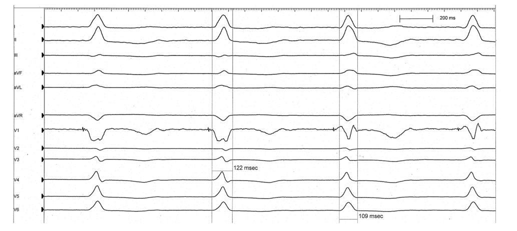
Figure 2. ECG of the QRS morphology change at a sweep speed of 100 mm/s

QUESTION

What does the change in morphology seen in figure 2 represent?

  • Failure to capture

  • Pseudo-fusion

  • Selective capture

  • Pseudo-pseudo-fusion

Answer
Selective capture

Explanation/Discussion

The ECGs here demonstrate the change from non-selective capture of the septal myocardium at high output to selective capture of the cardiac conduction system at a lower output.

Positioning the RV lead in the septum with the aim of pacing the left bundle branch of the cardiac conduction system allows for a more physiological method of cardiac pacing which produces synchronous contraction of the ventricles, first described in 2017 by Huang et al1, as opposed to the asynchronous contraction produced by pacing the right ventricular apex.

Selective capture means that only the conduction system is being captured, ‘hi-jacking’ the natural electrical pathway of the heart and producing a more physiological ECG. Whereas non-selective capture occurs when enough energy is delivered through the pacing lead to capture both the conduction system and the surrounding myocardium, resulting in a broader QRS complex with a right-sided septal morphology2.

References

  1. Huang, W., Su, L., Wu, S., Xu, L., Xiao, F., Zhou, X. and Ellenbogen, K.A. 2017. A Novel Pacing Strategy With Low and Stable Output: Pacing the Left Bundle Branch Immediately Beyond the Conduction Block. Canadian Journal of Cardiology. [Online]. 33(12), 1736.e1-1736.e3. Available from: http://dx.doi.org/10.1016/j.cjca.2017.09.013.
  2. Zhang, S., Zhou, X. and Gold, M.R. 2019. Left Bundle Branch Pacing. Journal of the American College of Cardiology. [Online]. 74(24), pp.3039–3049. Available from: http://dx.doi.org/10.1016/j.jacc.2019.10.039.

Disclaimer: The British Heart Rhythm Society (BHRS) collates submissions for the ECG/EGM challenge on this website. These submissions, along with any accompanying answers, are provided by external contributors and are published for informational purposes only. BHRS does not endorse, guarantee, or warrant the accuracy, completeness, or reliability of any submissions or answers provided.