May 2021

On-Call Remote Monitoring Disaster

Zara Patterson
Highly Specialised Cardiac Physiologist – Leeds Teaching Hospitals

Sunaina Asghar
Trainee Device Physiologist — Leeds Teaching Hospitals

Disclosure: The author has no conflict of interests to declare.

Background

A 78-year-old male underwent an elective battery replacement of an Abbott CRT-D. He has a background history of ischemic heart disease (IHD), NYHA class II heart failure (HF) symptoms, sinus rhythm with complete heart block and previous appropriate anti-tachycardia pacing (ATP) therapies for monomorphic ventricular tachycardia.

Implanted leads included;
RA Lead: Medtronic Select Secure Sense
RV Lead: SJM Durata 7120Q
LV Lead: SJM Quicksite 1058T

10 weeks post battery replacement an unscheduled remote alert was received for ‘RV lead impedance measurement out of range’. The right ventricular (RV) lead impedance trend is displayed in Figure 1.

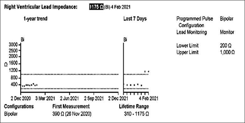
Figure 1 – RV lead impedance trend displaying recorded impedance measurements and range since elective battery replacement.

QUESTION 1

What action is required?

  • Plan for elective RV lead replacement

  • Schedule a remote check in one-month to review impedance trend

  • Investigate further with an in-person check

  • No further action required

Answer
Investigate further with an in-person check.

Explanation:

The lead impedance trend in Figure 1 shows a stable RV lead impedance at ~ 400 ohms. The most recent two measurements have suddenly risen above the programmed upper limit (1000 ohms) set in the device, triggering the remote alert. A sudden rise in impedance twice the previously recorded measurements should prompt further investigation  with an in-person device check as it could be an early indication and sign of lead failure.

Investigation

The patient was brought into clinic for further investigation. Device interrogation found all measurements to be satisfactory. Multiple RV lead impedance measurements were undertaken whilst performing isometric manoeuvres and during pocket manipulation with no evidence of high impedance values (all measurements stable at ~ 400 ohms), no ventricular lead noise, inhibition of pacing or loss of capture seen. A chest X-Ray was performed and compared to the post implant X-ray. There were no obvious lead pin/connector block abnormalities, lead fracture or lead irregularities seen.

As there was no clear evidence of RV lead failure and as the patient had access to remote monitoring, the patient was scheduled for a 1-month remote check and telephone consultation. The impedance upper limit alert range was left at 1000 ohms, to monitor for further impedance spikes. He was counselled on the importance of keeping his remote monitor plugged in and advised to contact the department if he experienced any symptoms of dizziness &/or loss of consciousness. The patient was also advised to contact the department if they had a shock from their device as per local protocol.

10 days following the face-to-face check…

10 days following the face to face check the on-call cardiac physiologist received another Merlin remote alert, this time for ‘ATP delivery and aborted shock’ Transmission details and relevant electrograms are displayed in Figures 2a-b.

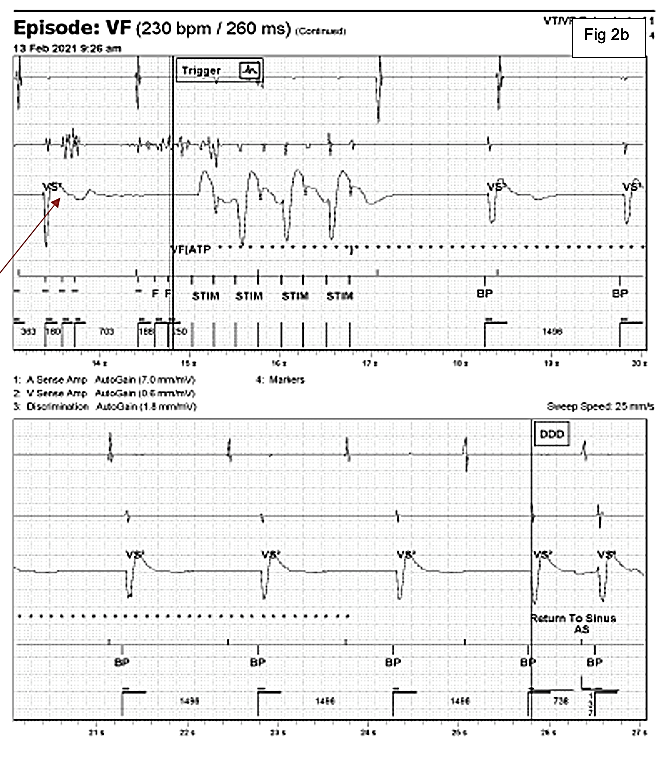
Figure 2a-b: Automatic Merlin.net alert transmission for ATP delivery and aborted shock. Channels displayed top to bottom, include the atrial EGM (A sense amp), right ventricular near field EGM (V sense amp), the far field discrimination channel and marker channel.

EGM Description

The EGM shows an episode of noise which receives an inappropriate ATP therapy. At the start of the trace in Figure 2a the EGM shows an atrial sense (As) biventricular paced (BP) rhythm. There is then a sudden change on the near field RV channel with high frequency noise signals detected which the device detects as ventricular events falling in the VF detection zone. These events are marked with an F for fibrillation sensing. At the end of the 12th F marker, ATP is delivered, marked as STIM on the trace. At the same time the device starts to charge (star markers). When the star markers are no longer displayed the device is charged ready to deliver a shock. The shock is then fortunately aborted with the disappearance of the high frequency noise signals on the near field channel and a return to sinus marker declares the end of the episode.

QUESTION 2

Why is ATP therapy delivered?

  • Short detection interval

  • Unsuccessful far field sensing

  • Committed therapy

  • Noise discriminator programmed off

Answer

Unsuccessful far field sensing

Explanation
Noise detected on the RV near field sensing circuit, is subsequently sensed and binned as VF (marked as an F and counted towards intervals to detection). At the point of detection (in this case 12 binned VF intervals), the Secure Sense RV Lead noise algorithm (designed to detect RV lead noise) was not applied as it had switched to passive function, due to a lack of sensing on the far-field channel. This is indicated on the trace by a single VS2 (shown by arrow in Figure 2b) marker during the detection period. The device perceives this single sensed beat as indicative of poor or inaccurate far-field sensing and therefore switches to passive. This is to reduce the chance of withholding therapy for a genuine ventricular arrhythmia. Therefore, the noise is detected as VF and results in delivery of inappropriate ATP and an aborted shock.

Management Strategy:
The history and recorded episode were highly suspicious of RV lead failure. As there was a risk of further inappropriate therapies and pacing inhibition the patient was immediately contacted and urgently admitted to our coronary care unit.

ICD therapies were disabled and re-programming attempted to avoid pacing inhibition. R wave sensitivity settings were assessed but it was not possible to avoid oversensing of noise by any reprogramming of the sensitivity threshold. Programming to DOO was considered however there was a high burden of ventricular ectopy and concerns of triggering an R on T episode. LV only pacing was also not an option for this device.

The best short term fix was to programme the mode to DDT to limit the inhibition during episodes of ventricular lead noise and the max trigger limit was decreased to 100 bpm to avoid pacing at excessively high rates with the patient monitored on telemetry overnight.

The next morning the patient was taken to the lab for RV lead replacement. Unfortunately, there was no left sided venous access with an occlusion at the left axillary vein therefore the procedure was abandoned. A decision was made to perform a full left sided system extraction and to implant a new CRT-D system on the right side. The extraction proved complex but was successful. Examination of the extracted RV lead found conductor externalisation proximal to the RV coil which was clearly the cause of the patients’ problems. A week later the patient had an uncomplicated right sided CRT-D implant with normalisation of the ECG. Follow up has been uneventful since implantation of the new device.

Learning Points
In conclusion, this case demonstrates the benefit of remote monitoring in the early identification of potential lead issues and adverse events for patients. In addition, the case was uncovered by the On-Call Physiologist demonstrating the merits of On-Call provision in centres dealing with complex device patients.

Despite a thorough in person review we were unable to reproduce noise episodes with manoeuvres reminding us that the location of the lead failure is a key determinant in whether noise can be reproduced.

As demonstrated in this case study, discrimination algorithms for noise can be complex and may not always be successful, knowledge of how these algorithms work is essential to understand device behaviour.

A final thought perhaps was that fluoroscopy may have been useful at uncovering the externalised conductor compared to a standard X-ray, although this is not often routine practice and based on the information available at the time, it is unlikely that we would have managed this patient differently.

References:
1. St Jude Medical, User Manual – Bradycardia and Tachycardia – Secure Sense Algorithm 2018