October 2023

Change in EP EGM – What is the cause?

Phil Matthews Highly Specialist Cardiac Physiologist, Golden Jubilee University National Hospital, UK

Disclosure: The author has no conflict of interests to declare.

Background

A 51-year-old male patient was referred for a Cryo Pulmonary Vein Isolation (PVI) ablation procedure due to documented paroxysmal Atrial Fibrillation (pAF). Previously he had noted palpitations for several years and after commencing Sotalol 80mg bd, he felt that he was in sinus rhythm the majority of the time although with occasional short-lived episodes of palpitations. Regarding his lifestyle, he is an ex-smoker after cessation 5 years prior, does not drink caffeine but does drink up to 6 pints of beer on weekend evenings. Echocardiogram showed a structurally normal heart with no significant left atrial dilatation. Medical history includes musculoskeletal back pain which is controlled by Tramadol prn and depression which the patient takes Fluoxetine 20mg od. Due to potential interactions between Fluoxetine and Sotalol causing prolonged QT segments, the patient was offered the option of alternative anti-arrhythmic medications or the option of catheter ablation.

Cryo PVI was undertaken with 2 catheters, a SJM Inquiry Coronary Sinus catheter and a Medtronic Achieve Pulmonary Vein catheter. During the first application of cryoablation treatment to the Left Superior Pulmonary Vein (LSPV), the following EGM (sweep speed 100mm/sec) was captured 50 seconds from onset of treatment and 5 seconds after balloon temperature reached -40C: –
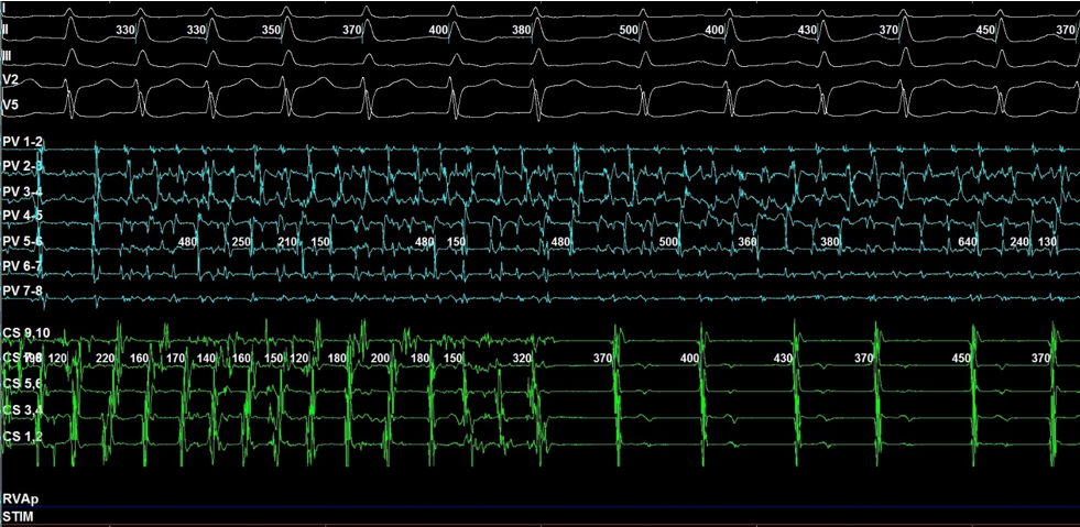
QUESTION

What can be seen in the captured EGM?  

  • Displacement of the Coronary Sinus catheter into Right Ventricle

  • Pulmonary Vein Exit Block

  • Onset of Accelerated Junctional Rhythm

  • Pulmonary Vein Entrance Block

Answer
Pulmonary Vein Exit Block

The above EGM is an example of Pulmonary Vein Exit Block, where electrical activity from the pulmonary vein cannot exit the vein into the left atrium.

By analysing the CS catheter, we can see that the electrical activity corresponds to the P wave on the EGM and shows concentric activation – from right (CS9-10) to left (CS1-2). The presence of P waves rules out Accelerated Junctional Rhythm, which would be evident with near simultaneous CS activity with the QRS.

In the first half of the EGM, we can see atrial fibrillation on the ECG and both the PV and CS catheter. After the 7th QRS complex, the ECG and CS catheter both show activity that corresponds to sinus rhythm; the PV catheter continues to show electrical activity that corresponds to AF. This shows that this particular PV – the LSPV – was the driver regarding this episode of AF and that electrical block has been achieved between the LSPV and the LA.

In 1998, Haïssaguerre et al. published a ground-breaking article that launched the concept of atrial fibrillation ablation. It identified that the pulmonary veins were an important source of atrial ectopics that initiate atrial fibrillation. Of the 69 identified foci, 65 (94%) were identified within the pulmonary veins. Of these 65 foci, 31 (48%) were located within the left superior; 11 (17%) in the left inferior; 17 (26%) in the right superior and 6 (9%) in the right inferior.

This gave rise to the concept of pulmonary vein isolation which is still in use today. The strategy of Wide Area Circumferential Ablation (WACA) of the PVs required the creation of transmural lesions inside the LA around the ostia of the PVs, guided by fluoroscopy and the usage of 3D mapping systems. Success rates varied and multiple procedures were frequently required to address areas of recovered LA-PV conduction. Additionally, the approach required a point-by-point fashion which was time-consuming and technically challenging with difficulties in creating durable transmural lesions.

In 2016 the FIRE AND ICE trial assessed whether the use of cryoballoon ablation was non-inferior to radiofrequency (RF) ablation in patients with drug-refractory pAF. Cryoablation utilises a balloon that occludes the pulmonary vein and cools the tissue using a liquid refrigerant with a target temperature between -40 and -60C. This creates intra and extracellular ice crystals to form which lead to cellular death and eventual scarring and fibrosis. The trial showed that cryoablation was non-inferior to RF with regards of efficacy and safety, with shorter procedure and left atrial dwell times compared to RF. To this day it remains a cornerstone in the treatment of AF, however, on the horizon is the evolving field of pulsed field ablation with promising emerging clinical data.

References

  1. Haïssaguerre M, et al. Spontaneous initiation of atrial fibrillation by ectopic beats originating in the pulmonary veins. N Engl J Med 1998 Sep 3;339(10):659-66
  1. Kuck K-H, et al. Cryoballoon or Radiofrequency Ablation for Paroxysmal Atrial Fibrillation. N Engl J Med 2016; 374:2235-2245

Disclaimer: The British Heart Rhythm Society (BHRS) collates submissions for the ECG/EGM challenge on this website. These submissions, along with any accompanying answers, are provided by external contributors and are published for informational purposes only. BHRS does not endorse, guarantee, or warrant the accuracy, completeness, or reliability of any submissions or answers provided.